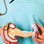
Hidden Threat: Pancreatic Cysts
Despite their seemingly harmless nature, pancreatic cysts can pose a serious danger. While many of them do not threaten health, some types, such as mucinous cysts and intraductal papillary mucinous neoplasms (IPMNs), can transform into malignant formations. Modern diagnostic methods and a comprehensive approach to treatment available at the Ichilov Clinic allow for timely detection of these pathologies and prevention of the development of oncological diseases.
⚠️ When a Pancreatic Cyst Becomes Dangerous
- ➤ Size exceeds 3 cm — increased risk of malignancy;
- ➤ Rapid growth of the cyst — an increase of 5 mm or more in a year requires urgent examination;
- ➤ Thickening of the cyst walls or the appearance of septa — a sign of possible degeneration;
- ➤ Expansion of the main pancreatic duct over 5 mm — may indicate obstruction by a tumour;
- ➤ Appearance of symptoms — abdominal pain, jaundice, weight loss, nausea, vomiting;
- ➤ Increased tumor markers (CA 19-9, CEA) in the cyst contents during biopsy.
At the Ichilov Clinic, each case of pancreatic cyst is reviewed by a panel of doctors: a gastroenterologist, surgeon, oncologist, and radiologist jointly determine the strategy — observation or surgery.
According to studies, pancreatic cysts are diagnosed in approximately 5% of patients undergoing computed tomography (CT) of the abdominal organs. In most cases, they do not cause discomfort and do not require treatment. However, cysts with high oncogenic potential represent a serious medical problem. That is why they are often referred to as a “ticking time bomb,” as untimely intervention can lead to irreversible consequences.
Diagnostic Accuracy – Key to Successful Treatment
Thanks to innovative imaging technologies available at the Ichilov Clinic, such as high-precision CT and magnetic resonance imaging (MRI), doctors have the opportunity to detect pancreatic cysts at the earliest stages. This significantly increases the chances of successful treatment and a favorable prognosis for patients.
In Israel, a comprehensive interdisciplinary approach is used for the diagnosis of cysts, combining the efforts of gastroenterologists, radiologists, and surgeons. The standard diagnostic algorithm includes:
- ➤ CT and MRI: allow detailed visualization of the cyst, determining its size, structure, and location.
- ➤ Endoscopic ultrasound (EUS): provides more accurate information about the characteristics of the cyst and, if necessary, allows for biopsy.
- ➤ Laboratory tests: help assess the overall condition of the patient and the likelihood of a malignant process.
Correct Approach to Treatment
The choice of treatment method depends on the type, size, and location of the cyst, as well as the overall condition of the patient. Israeli clinics offer a wide range of treatment methods, including:
- ➤ Dynamic observation is applied for safe and stable cysts.
- ➤ Surgical treatment is indicated in cases of suspected malignant degeneration or disease progression.
Modern surgery offers minimally invasive techniques, such as laparoscopy and robotic surgeries, which significantly reduce the rehabilitation period and lower the risk of complications.
Cyst Diagnosis Program at Ichilov
Day 1 — Initial Consultation and Imaging:
- ➤ Gastroenterologist appointment — review of medical history, symptoms, previous examinations;
- ➤ Abdominal CT with contrast — assessment of the size, structure, and location of the cyst;
- ➤ MRI of the pancreas with MRCP (magnetic resonance cholangiopancreatography) — detailed visualization of the ducts;
- ➤ Blood tests: general, biochemistry, tumor markers (CA 19-9, CEA), amylase, lipase.
Day 2 — Endoscopic Diagnosis:
- ➤ EUS (endoscopic ultrasound) — high-precision visualization of the cyst “from the inside” through the stomach or duodenum;
- ➤ Fine-needle biopsy of the cyst under EUS guidance — collection of contents for cytological and biochemical analysis;
- ➤ Analysis of fluid from the cyst: amylase level (to differentiate pseudocysts from true cysts), tumor markers (CEA >192 ng/ml indicates mucinous nature), cytology (search for atypical cells).
Day 3 — Consultation and Treatment Plan:
- ➤ Interdisciplinary consultation: gastroenterologist, oncologist, abdominal surgeon, radiologist;
- ➤ Discussion of the results of all examinations;
- ➤ Determination of the type of cyst and degree of oncological risk;
- ➤ Choice of strategy: dynamic observation (with a schedule for follow-up examinations) or surgical treatment;
- ➤ If surgery is necessary — consultation with the surgeon, choice of method (laparoscopy, robotic surgery, open resection).
Duration of examination: 3 days. Results with conclusions in Russian. If surgery is required — hospitalization may be possible the following week.
Patient Story
Recently, a patient came to the clinic who was found to have a pancreatic cyst during a routine screening, which posed a serious threat. After assessing the risks, a decision was made for surgical intervention. Histological examination confirmed the concerns — the cyst was on the verge of degeneration into cancer. Thanks to timely surgery, the patient did not have to undergo chemotherapy.
Pancreatic cysts are a serious condition requiring professional medical intervention. Timely seeking medical help can save lives.